Photsymbiosis in Spotted Salamander Eggs

The green alga Oophila amblystomatis grows inside the egg capsules of the spotted salamander, and in some cells, inside the embryo itself. It is the only known case of a photosynthetic cell living inside the tissues of a vertebrate.
The problem
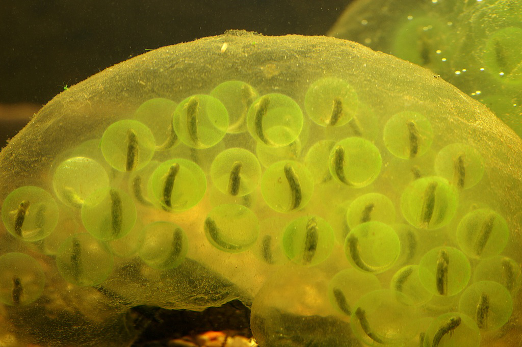
The first observation of green salamander eggs in the scientific literature is from the naturalist Henry Orr in 1888. Orr wrote that the developing eggs of Amblystoma [sic] “seem to present a remarkable case of symbiosis,” and went on: “I have not discovered how the algae enter the membrane, nor what physiological effect they have on the respiration of the embryo, but it seems probable that in this latter respect they may have an important influence.” Nearly 140 years later we understand a good deal about the mechanisms and dynamics of the salamander-alga symbiosis, and Orr’s first question stands: we still do not know how the algae get into the eggs.
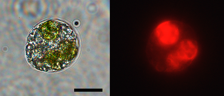
In 2011, Ryan Kerney and colleagues found that the egg-colonizing algae also enter embryonic tissues and individual cells. This is the only such case recorded in a vertebrate, and it is the exception that proves the rule: the algae are tolerated in an embryo, before adaptive immunity is established. That constraint holds, because at embryonic stages the host salamander’s immune system is not yet developed.
History
In the 1940s, seminal work by Perry Gilbert established the mutualism: embryos developing with algae hatch earlier, larger, and more successfully than those without.
In 1958, Hutchison and Hammen measured oxygen utilization in the association, and in 1962 the same pair followed carbon fixation in the egg (Hammen and Hutchison, Life Sciences 1:527–532). The carbon paper concluded that the association was not simply algal fixation followed by transfer to the embryo. They found instead that the embryos fix a considerable amount of inorganic carbon themselves. We reconfirmed and extended this in 2020, controlling for heterotrophic carbon fixation by the embryo and finding competition for carbon dioxide within the egg at early developmental stages.
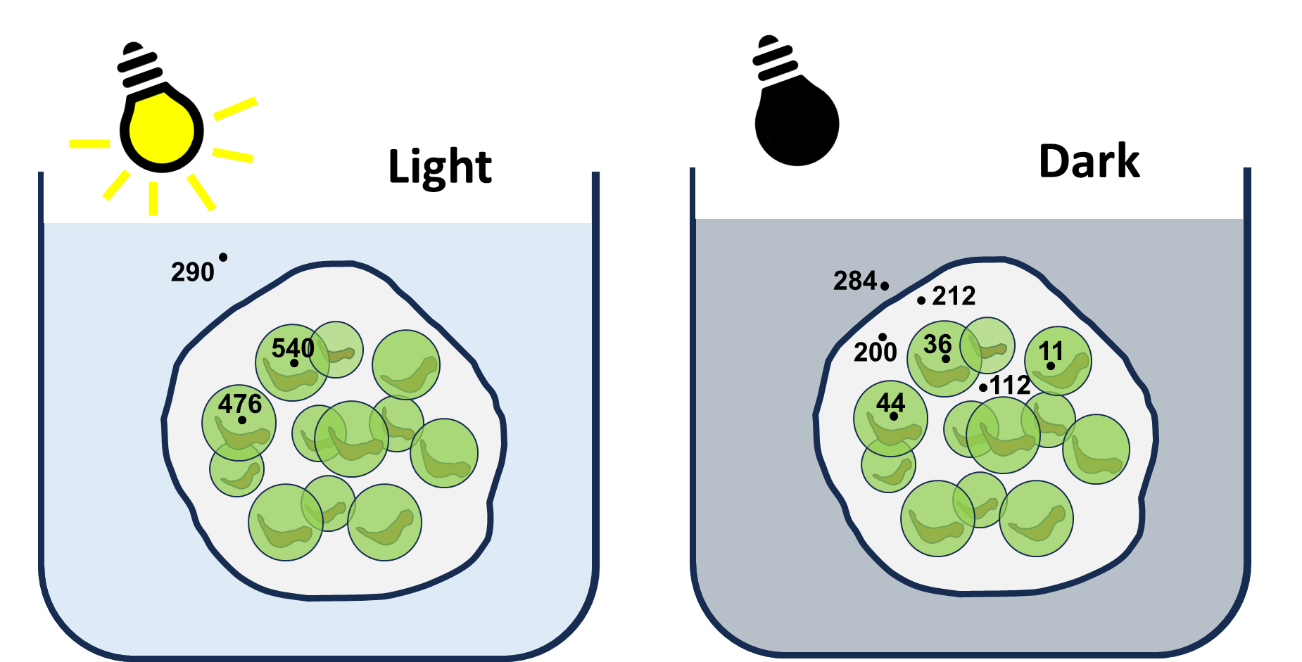
In 1986, Bachmann, Carlton, Burkholder, and Wetzel put an oxygen electrode inside the eggs themselves. In darkness oxygen was severely depleted, so whatever diffusion delivers through the jelly is not enough. In light it rose fast, past saturation, even when the water around the mass was close to anoxic. The oxygen an embryo has comes from photosynthesis, not from the pond.
In 1994, Pinder and Friet showed why the symbiosis matters so much to salamander embryos. Unlike wood frog egg masses, which host the same alga but are loose enough for water to circulate between eggs, spotted salamander masses are firm with no convective route at all. Diffusion cannot supply oxygen to the embryos in the middle, especially late in development, and the algae make up the difference. They also documented the daily swing: the egg mass goes hyperoxic in the light, and at night both algae and embryo consume oxygen, driving the eggs to a low oxygen state.
The 2011 finding by Kerney and colleagues reignited interest in the association.
In 2014, Small, Bennett, and Bishop observed ammonia uptake by algae in egg masses. In addition to supplying oxygen, the algae remove waste products that would otherwise build up to toxic levels in the diffusion-limited egg.
Also in 2014, Kim and colleagues mapped the phylogenetic relationships of Oophila, showing that the egg-associated algae form a clade across diverse amphibian hosts. Algae from the same group have since been identified in salamanders in Japan and frogs in Europe.
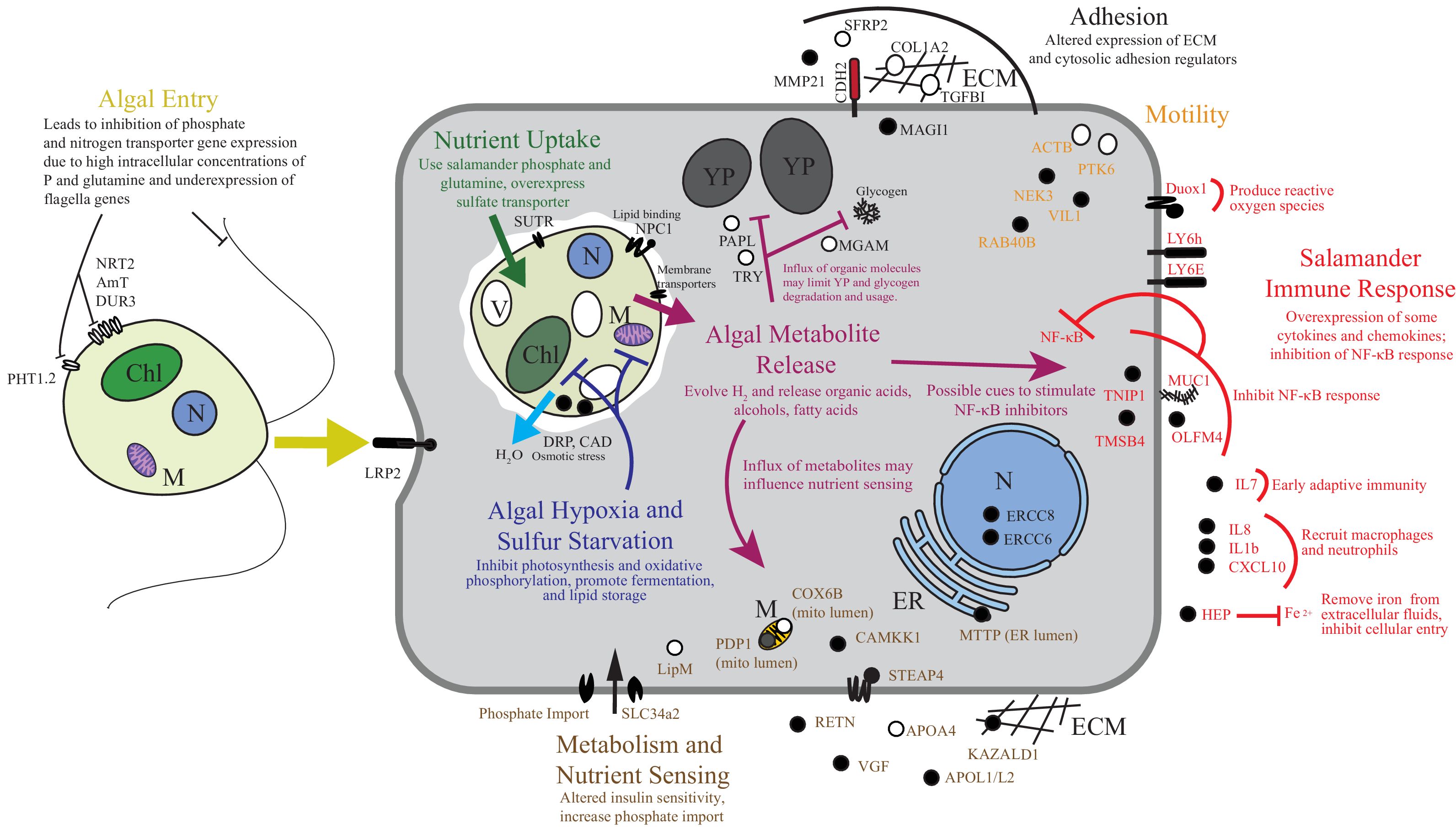
In 2017, we looked at transcriptional regulation during the endosymbiosis and found that the endosymbiotic algae are stressed and fermenting, while the host cells show an altered but muted immune signal and otherwise appear to tolerate them well.
Where it stands
Work continues on population structure and phylogenetics in the alga, and on genetic tools for working with it. We are currently doing metabolomics and transcriptomics on the system in collaboration with others. The system suits both questions of its own and broader ones about symbiosis, immunity, and how a cell finds its way through a crowded fluid.
Collaborators
- Ryan Kerney, Gettysburg College. Salamander development.
- Eunsoo Kim, Ewha Womans University. Protist phylogenetics and cell biology.
- Cory Bishop, St. Francis Xavier University. Embryo physiology and the egg capsule environment.
- Baptiste Genot, University of Tokyo. Cell biology of the symbiosis.
- Wendy Strangman and Robert Williamson, University of North Carolina Wilmington. Natural products chemistry.
- Vera Nikitashina and Georg Pohnert, Friedrich Schiller University Jena. Metabolomics.
- Gaëlle Toullec and Johan Decelle, Université Grenoble Alpes. Subcellular imaging, through the AtlaSymbio project.
Support
Current support
Currently seeking support for projects in this system.
Prior support

The Gordon and Betty Moore Foundation supported much of our work on this system.

The National Science Foundation funded my initial involvement through award 1428065.
An award from the William Procter Scientific Innovation Fund let us begin exploring natural products and immune interactions in the symbiosis.